|
Home » Health » Aluminum » Aluminum: Health Risks
Aluminum: Health Risks
atomic no. 13, atomic wt. 26.98, metal, row 4, col.3A, val. 3, orbits
2-8-3
Aluminum. Al; at. wt. 26.98; at. no. 13; valence 3. Discovered by Wöhler
in 1827. Obtained from cryolite (double sod. and aluminum fluoride) or
bauxite (native aluminum hydroxide) by electrolysis in electric
furnace.
Tin-white, malleable, ductile metal, with somewhat bluish tint; capable
of taking brilliant polish which is retained in dry air. In moist air
gradually oxidizes superficially. Available in bars, leaf, powder, sheets
or wire. d. 2.70. m. 660°. b. 1800°. Does not vaporize even at high temps.,
but finely divided aluminum dust is easily ignited, and may cause
explosions. Soluble in dil. HCl, H2SO4, in soln. KOH
and NAOH with evolution of hydrogen; almost insoluble in HNO3 or
acetic acid when hot.
Reduces the cations of many heavy metals to the metallic state. Solns.
of the metal in dil. HCl or neutral or slightly acid solns. of most
aluminum salts, yield with Na2S a white ppt. soluble in excess
of Na2S. Dil. neutral soln. of aluminum salts yield white
gelatinous ppt. on boiling with sod. acetate.
Use: As the pure metal or as alloys (magnalium, aluminum bronze, etc.)
for aircraft, utensils, apparatus, electrical conductors; instead of copper
in dental alloys. The coarse powder is used in aluminothermics (thermite
process); the fine powder as flashlight in Photography, in explosives,
fireworks and in aluminum paints; for absorbing occluded gases in manuf. of
steel. In testing for Au, As, Hg; coagulating colloidal solns. of As or Sb;
pptg. Cu; reducer for determining nitrates and nitrites; instead of Zn for
generating hydrogen in testing for As.
Grades available: Reagent, technical.
Med. Use: Inhalation of finely divided aluminum dust proposed as a means
of "binding" silica to prevent and reverse lung changes caused by silica
dust.
Aluminum Toxicity
"From the earliest days of food
regulation, the use of alum (aluminum sulphate) in foods has been
condemned. It is universally acknowledged as a poison in all countries. If
the Bureau of Chemistry had been permitted to enforce the law ... no food
product in the country would have any trace of ... any aluminum or
saccarin. No soft drink would contain caffeine or hebromin; no bleached
flour would be in interstate commerce. Our food and drugs would be wholly
without adulteration ... and the health of our people would be vastly
improved and their life greatly extended."
From History of crime against the Food Laws (1929) by Dr. Wiley, the
prime mover behind the original Pure Food Law and Director of the FDA. He
resigned in disgust in 1912 over exceptions granted to the law and lack of
enforcement.
Aluminum has been exempted from tesitng for safety by the FDA under a
convoluted logic wherein it is classified as GRAS. (Generally Regarded As
Safe.) It has never been tested by the FDA on its safety and there are NO
restrictions whatever on the amount or use of aluminum.
There are over 2000 references in the National Library of Medicine on
adverse effects of alumium. The following were extracted to provide a small
sample of the range of toxicity of aluminum.
Chemical Registry
Aluminum toxicity has been recognized in many settings where exposure is
heavy or prolonged, where renal function is limited, or where apreviously
accumulated bone burden is released in stress or illness. Toxicity may
include: encephalopathy (stuttering, gait disturbance, myoclonic jerks,
seizures, coma, abnormal EEG) osteomalacia or aplastic bone disease (
associated with painful spontaneous fractures, hypercalcemia, tumorous
calcinosis ) proximal myopathy, increased risk of infection, increased left
ventricular mass and decreased myocardial function microcytic anemia with
very high levels, sudden death.
Aluminum is ubiquitous in our environment; it is the third most
prevalent element in the earth's crust. The gastrointestinal tract is
relatively impervious to aluminum, absorption normally being only about 2%.
Aluminum is absorbed by a mechanism related to that for calcium. Gastric
acidity and oral citrate favors absorption, and H2-blockers reduce
absorption. As is true for several trace elements, transferrin is the
primary protein binder and carrier for aluminum in the plasma, where 80% is
protein bound and 20% is free or complexed to small molecules such as
citrate.
Cells appear to take up aluminum from transferrin rather than from
citrate. Purified preparations of ferritin from brain and liver have been
found to contain aluminum.
It is not known if ferritin has a specific binding site for aluminum.
Factors regulating the migration of aluminum across the blood–brain barrier
are not well understood.
Serum aluminum correlates with encephalopathy; red cell aluminum
correlates with microcytic anemia, and bone aluminum correlates with
aluminum bone disease.
Basal PTH when elevated appears to protect bone and thereby favor CNS
toxicity.
Other factors favoring one form of toxicity over another are not well
understood.
Aluminum toxicity has been reported to impair the formation and release
of parathyroid hormone. The parathyroid glands concentrate aluminum above
levels in surrounding tissues. Treatment of aluminum toxicity in renal
failure patients often reactivates hyperparathyroidism, which to a certain
extent is helpful for bone remodeling and healing.
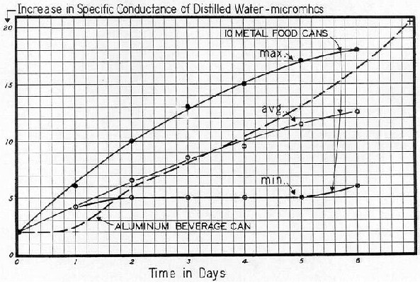
Distilled water was placed in metal containers and the amount of the
"Metal Can" that disolved into the distilled water was measured daily
using Specific Conductance readings. You can divide the SC number by 2
to get the approxamite amount of atoms in ppm
( mg / l ).
4 ppm of aluminum in human blood can cause it to colagulate.
Aluminum in humans is documented to Inhibit Learning.
Aluminum neurotoxicity in
preterm infants receiving intravenous-feeding solutions.
Bishop N.J. – Morley R. – Day J.P. – Lucas A.
From: N Engl J Med (1997 May
29) 336(22):1557-61
Aluminum, a contaminant of commercial intravenous–feeding solutions, is
potentially neurotoxic. We investigated the effect of perinatal exposure to
intravenous aluminum on the neurologic development of infants born
prematurely.
RESULTS: The 90 infants who received the standard feeding solutions had
a mean (± SD) Bayley Mental Development Index of 95 ±22, as compared with
98 ±20 for the 92 infants who received the aluminum-depleted solutions
(P=0.39). The former were significantly more likely (39 percent, vs. 17
percent of the latter group; P=0.03) to have a Mental Development Index of
less than 85, increasing their risk of subsequent educational problems. For
all 157 infants without neuromotor impairment, increasing aluminum exposure
was associated with a reduction in the Mental Development Index (P=0.03),
with an adjusted loss of one point per day of intravenous feeding for
infants receiving the standard solutions. In preterm infants, prolonged
intravenous feeding with solutions containing aluminum is associated with
impaired neurologic development.
Aluminum-containing emboli in
infants treated with extracorporeal membrane oxygenation.
Vogler C. – Sotelo-Avila C. – Lagunoff D. – Braun P. – Schreifels J.A. –
Weber T.
From: N Engl J Med (1988 Jul
14) 319(2):75-9
We found fibrin thrombi or thromboemboli at autopsy in 22 of 23 infants
with respiratory failure who had been treated with venoarterial
extracorporeal membrane oxygenation (ECMO). In addition, distinctive
basophilic aluminum-containing emboli were found in 12 of the infants; the
distribution of these emboli was similar to that of the thromboemboli,
except that an aluminum-containing embolus was found in a lung in only 1
infant. Sixteen infants had pulmonary thrombi or thromboemboli. We also
found friable aluminum-containing concretions adhering loosely to the
mixing rods of heat exchangers that had been used to warm the blood flowing
through the ECMO circuit; such concretions were not present on unused
mixing rods. We propose that these aluminum-containing concretions
developed as the silicone coating of the heat exchanger wore away and
aluminum metal was exposed to warm, oxygenated blood and that fragments of
aluminum-containing concretions formed emboli. This hypothesis is supported
by the fact that aluminum-containing emboli were generally not present in
the lungs, which are bypassed by ECMO.
Sequential serum aluminum and
urine aluminum: creatinine ratio and tissue aluminum loading in infants
with fractures/rickets.
Koo W.W. – Krug-Wispe S.K. – Succop P. – Bendon R. – Kaplan L.A.
From: Pediatrics (1992 May)
89(5 Pt 1):877-81
Aluminum toxicity is associated with the development of bone disorders,
including fractures, osteopenia, and osteomalacia. Fifty-one infants with a
mean (± SEM) birth weight of 1007 ±34 g, gestational age of 28.5 +/-0.3
weeks, and serial radiographic documentation at 3, 6, 9, and 12 months for
the presence (n = 16) or absence (n = 35) of fractures and/or rickets were
studied at the same intervals to determine the serial changes in serum
aluminum concentrations and urine aluminum-creatinine ratios. Autopsy bone
samples were used to determine the presence of tissue aluminum. One infant
who received aluminum-containing antacid had marked increase in serum
aluminum to 83 micrograms/L while urine aluminum-creatinine ratio increased
from 0.09 to a peak of 8.53. Vertebrae from three infants at autopsy (full
enteral feeding was tolerated for 37 and 41 days in two infants,
respectively) showed aluminum deposition in the zone of provisional
calcification and along the newly formed trabecula.
Aluminum in parenteral
solutions revisited — again.
Klein G.L.
From: Am J Clin Nutr (1995
Mar) 61(3):449-56
It has been a dozen years since aluminum was first shown to contaminate
parenteral nutrition solutions and to be a contributing factor in the
pathogenesis of metabolic bone disease in parenteral nutrition patients as
well as in uremic patients. However, there are no regulations in place to
effectively reduce aluminum contamination of various parenterally
administered nutrients, drugs, and biologic products. The purpose of this
review is fourfold: 1.) to summarize our knowledge of the adverse effects
of aluminum on bone formation and mineralization in parenteral nutrition
patients; 2.) to discuss the possible role of aluminum in the osteopenic
bone disease of preterm infants; 3.) to show how lack of regulations
covering aluminum content of parenteral solutions can lead to vulnerability
of new groups of patients to aluminum toxicity, the example being given
here is that of burn patients
Aluminum-induced anemia.
From: Am J Kidney Dis (1985 Nov) 6(5):348-52
... many questions still remain unanswered, it is clear that aluminum
causes a microcytic hypoproliferative anemia and is a factor responsible
for worsening anemia in patients with end-stage renal disease.
Arch Dermatol (1984 Oct) 120(10):1318-22
Three patients had subcutaneous nodules at the sites of previous
injections of vaccine containing tetanus toxoid, showed aluminum crystals
in the nodules from two patients. From the evidence available, we believe
that these nodules are a complication of inoculations with
aluminum-containing vaccines.
Persistent subcutaneous nodules
in patients hyposensitized with aluminum-containing allergen extracts.
Garcia-Patos V. – Pujol R.M. – Alomar A. – Cistero A. – Curell R. –
Fernandez-Figueras M.T. – de Moragas J.M.
From: Arch Dermatol (1995
Dec) 131(12):1421-4
These lesions have been mainly attributed to a hypersensitivity reaction
to aluminum hydroxide, which is used as an absorbing agent in many vaccines
and hyposensitization preparations. Patch tests with standard antigens and
aluminum compounds and histopathologic and ultrastructural studies were
performed on 10 patients with persistent subcutaneous nodules on the upper
part of their arms after injection of aluminum-adsorbed dust and/or pollen
extracts. The nodules appeared 1 month to 6.5 years after injections.
Trace metals and degenerative
diseases of the skeleton.
Savory J. – Bertholf R.L. – Wills M.R.
From: Acta Pharmacol Toxicol
(Copenh) (1986) 59 Suppl 7:282-8
Aluminum related osteodystrophy is the most important manifestation of
trace metal toxicity related to degenerative diseases of the skeleton.
Postvaccinal sarcomas in the
cat: epidemiology and electron probe microanalytical identification of
aluminum.
Hendrick M.J. – Goldschmidt M.H. – Shofer F.S. – Wang Y.Y. – Somlyo
A.P.
From: Cancer Res (1992 Oct
1) 52(19):5391-4
An increase in fibrosarcomas in a biopsy population of cats in the
Pennsylvania area appears to be related to the increased vaccination of
cats following enactment of a mandatory rabies vaccination law.
The majority of fibrosarcomas arose in sites routinely used by
veterinarians for vaccination, and 42 of 198 tumors were surrounded by
lymphocytes and macrophages containing foreign material identical to that
previously described in postvaccinal inflammatory injection site reactions.
Some of the vaccines used have aluminum-based adjuvants, and macrophages
surrounding three tumors contained aluminum oxide identified by electron
probe microanalysis and imaged by energy-filtered electron microscopy.
Persistence of inflammatory and immunological reactions associated with
aluminum may predispose the cat to a derangement of its fibrous connective
tissue repair response, leading to neoplasia.
Aspects of aluminum toxicity.
Hewitt C.D. – Savory J. – Wills M.R.
From: Clin Lab Med (1990
Jun) 10(2):403-22
Attention was first drawn to the potential role of aluminum as a toxic
metal over 50 years ago, but was dismissed as a toxic agent as recently as
15 years ago. The accumulation of aluminum, in some patients with chronic
renal failure, is associated with the development of toxic phenomena;
dialysis encephalopathy, osteomalacic dialysis osteodystrophy, and an
anemia. Aluminum accumulation also occurs in patients who are not on
dialysis, predominantly infants and children with immature or impaired
renal function. Aluminum has also been implicated as a toxic agent in the
etiology of Alzheimer's disease, Guamiam amyotrophic lateral sclerosis, and
parkinsonism-dementia.
Soft tissue sarcoma associated
with aluminum oxide ceramic total hip arthroplasty. A case report.
Ryu R.K. – Bovill E.G. Jr – Skinner H.B. – Murray W.R.
From: Clin Orthop (1987
Mar)(216):207-12
Malignant tumors around fracture fixation implants have been reported
sporadically for many years. Recently, however, reports of sarcomatous
degeneration around a standard cemented hip arthroplasty and around
cobalt-chromium-bearing hip arthroplasties raise new questions of the
malignant potential of metallic ends prostheses. Sarcomatous changes around
aluminum oxide ceramics seem not to have been reported in the literature.
The present report may be the first documented case of an aggressive soft
tissue sarcoma detected 15 months after the patient had an uncemented
ceramic total hip arthroplasty. If a causal relationship exists, the
incidence of this phenomenon in the United States is 250 times greater than
would be expected from statistics on soft tissue sarcoma at the hip.
Aluminum-induced granulomas in
a tattoo.
McFadden N. – Lyberg T. – Hensten-Pettersen A.
From: J Am Acad Dermatol
(1989 May) 20(5 Pt 2):903-8
Aluminum was the only nonorganic element present in the test site
tissue. This is the first report of confirmed aluminum-induced,
delayed-hypersensitivity granulomas in a tattoo.
Delayed healing in
full-thickness wounds treated with aluminum chloride solution. A histologic
study with evaporimetry correlation.
Sawchuk W.S. – Friedman K.J. – Manning T. – Pinnell S.R.
From: J Am Acad Dermatol
(1986 Nov) 15(5 Pt 1):982-9
Wounds were treated either with 30% aluminum chloride solution or ferric
subsulfate solution or were allowed to clot with minimal pressure from a
gauze pad. Delay in reepithelialization was noted histologically both in
wounds treated with aluminum chloride and in those treated with ferric
subsulfate compared to controls. Presumably this delay was the result of
tissue necrosis caused by these hemostatic agents, resulting in slightly
larger and less cosmetically acceptable scars. Plots of evaporimetry data
revealed a biphasic pattern of water loss during healing, with an initial
rapid decline in water loss followed by a much slower decline.
Aluminium and injection site
reactions.
Culora G.A. – Ramsay A.D. – Theaker J.M.
From: J Clin Pathol (1996
Oct) 49(10):844-7
To alert pathologists to the spectrum of histological appearances that
may be seen in injection site reactions related to aluminium, showed
unusual features not described previously. In one, there was a sclerosing
lipogranuloma-like reaction with unlined cystic spaces containing
crystalline material. The other case presented as a large symptomatic
subcutaneous swelling which icroscopically showed diffuse and wide-spread
involvement of the subcutis by a lymphoid infiltrate with prominent
lymphoid follicles.
CONCLUSIONS: This report highlights the changes encountered in aluminium
injection site reactions and emphasises that the lesions have a wider range
of histological appearances than described previously.
Aluminum and gallium arrest
formation of cerebrospinal fluid by the mechanism of OH- depletion.
Vogh B.P. – Godman D.R. – Maren T.H.
From: J Pharmacol Exp Ther
(1985 Jun) 233(3):715-21
AlCl3 or GaCl3 was added to artificial
cerebrospinal fluid and perfused through the cerebral ventricles of the
rat. Depending on the metal and its concentration (1-10 mM) the pH of the
perfusate ranged from 7.2 to 3.5. At 10 mM metal chloride, yielding pH 4.7
(Al) or 3.5 (Ga), formation of cerebrospinal fluid was suppressed 100%.
This mechanism may also account for the antiperspirant action of Al
salts.
Aluminum toxicity and albumin.
Kelly A.T. – Short B.L. – Rains T.C. – May J.C. – Progar J.J.
From: ASAIO Trans (1989
Jul-Sep) 35(3):674-6
During a study of priming solutions for extracorporeal membrane
oxygenation (ECMO) in the intensive care nursery, it was discovered that
those solutions using certain brands of 25% albumin contained aluminum
levels within the toxic range. When the brand was changed to a brand known
to have a lower aluminum (Al) content, a marked drop in priming solution Al
levels was measured.
The role of aluminium for
adverse reactions and immunogenicity of diphtheria-tetanus booster vaccine.
Mark A. – Granstrom M.
From: Acta Paediatr (1994
Feb) 83(2):159-63
235 schoolchildren aged 10 years received either a regular,
aluminium-adsorbed diphtheria-tetanus vaccine or the same vaccine in fluid
form, in order to investigate if local side effects could be diminished by
exclusion of aluminium. System reactions were rare and local reactions
frequent in both groups but larger local reactions were even more
pronounced in the non-adsorbed vaccine group.
Potroom palsy? Neurologic
disorder in three aluminum smelter workers.
Heyer N.J.
From: Arch Intern Med (1985
Nov) 145(11):1972-5
We studied three patients with a progressive neurologic disorder, all of
whom had worked for over 12 years in the same potroom of an aluminum
smelting plant. All had incoordination and an intention tremor. Two of the
three patients had cognitive deficits, and the most severely affected
patient also had spastic paraparesis. None had involvement of the
peripheral nervous system. Despite extensive evaluations, the cause of
these patients' problems remains obscure.
Neurotoxic effects of aluminum in animals are directed at the central
nervous system, and theoretically long-term low-level exposure to aluminum
in the potroom could explain the findings in our patients.
Reducing aluminum: an
occupation possibly associated with bladder cancer
Theriault G. – De Guire L. – Cordier S.
From: Can Med Assoc J (1981)
124(4):419-422,425
These findings suggest that employment in an aluminum reduction plant
accounts for part of the excess of bladder cancer in the region studied.
(Author abstract) (85 Refs)
Immunohistochemical study of
microtubule-associated protein 2 and ubiquitin in chronically
aluminum-intoxicated rabbit brain.
Takeda M. – Tatebayashi Y. – Tanimukai S. – Nakamura Y. – Tanaka T. –
Nishimura T.
From: Acta Neuropathol
(Berl) (1991) 82(5):346-52
Experimental neurofibrillary change was produced in rabbit brain by
daily subcutaneous aluminum tartrate injection for 40 days.
Neurotoxic effects of aluminium
on embryonic chick brain cultures.
From: Acta Neuropathol (Berl) (1994) 88(4):359-66
Toxic damage of brain cells by aluminium (Al) is discussed as a possible
factor in the development of neurodegenerative disorders in humans. Effects
of Al on cell viability (lysosomal and mitochondrial activity) and
differentiation (synthesis of cell-specific proteins) were found to the
brain area specific with the highest sensitivity observed in optic
tectum.
Aluminium in tooth pastes and
Alzheimer's disease.
Verbeeck R.M. – Driessens F.C. – Rotgans J.
From: Acta Stomatol Belg
(1990 Jun) 87(2):141-4
The role of aluminium from tooth pastes may be even more important than
that from the drinking water.
Persistent subcutaneous nodules
in children hyposensitized with aluminium-containing allergen extracts.
Frost L. – Johansen P. – Pedersen S. – Veien N. – Ostergaard P.A. –
Nielsen M.H.
From: Allergy (1985 Jul)
40(5):368-72
A follow-up study of 202 children who had received hyposensitization
with aluminium-containingallergens showed that 1-3 years after cessation of
hyposensitization 13 children still had severely treatment-resistant
subcutaneous nodules in their forearms. Because of their long persistence
the nodules of six children were studied in detail. Histologically, the
nodules showed infiltration with lymphocytes (forming germinal centres),
macrophages, plasma cells, mast cells and a few eosinophils.
In five patients aluminium crystals were found scattered between the
cells and, in addition, the phagosomes of the macrophages contained
aluminium. Patch tests for aluminium were positive in four of the six
patients.
Contact sensitivity to
aluminium in a patient hyposensitized with aluminium precipitated grass
pollen.
Clemmensen O. – Knudsen H.E.
From: Contact Dermatitis
(1980 Aug) 6(5):305-8
Standard patch testing of a patient with eczema revealed positive
reactions to the aluminium discs used for testing.
Behavioural effects of
gestational exposure to aluminium.
Rankin J. – Sedowofia K. – Clayton R. – Manning A.
From: Ann Ist Super Sanita
(1993) 29(1):147-52
The involvement of aluminium in the aetiology of a number of human
pathological diseases has altered its status from being a nontoxic,
nonabsorbable, harmless element. This maybe of particular concern to the
developing foetus which is more susceptible to agents and at lower levels
than the adult. Little attention has been given to aluminium's potential
reproductive toxicity until recently and further research is required for a
full evaluation of its toxicity. Our preliminary results demonstrate
behavioural and neurochemical alterations in the offspring of mice exposed
to aluminium during gestation. Further, the effects of such exposure are
also present in the adult animal suggesting persistent changes in behaviour
following prenatal exposure.
The absence of extracellular
calcium potentiates the killing of cultured hepatocytes by aluminum
maltolate.
Snyder J.W. – Serroni A. – Savory J. – Farber J.L.
From: Arch Biochem Biophys
(1995 Jan 10) 316(1):434-42
This data defines a new model in which aluminum kills liver cells by a
mechanisms distinct from previously recognized pathways of lethal cell
injury. It is hypothesized that aluminum binds to cytoskeletal proteins
intimately associated with the plasma membrane. This interaction eventually
disrupts the permeability barrier function of the cell membrane, an event
that heralds the death of the hepatocyte.
Sensitization to aluminium by
aluminium-precipitated dust and pollen extracts.
Castelain P.Y. – Castelain M. – Vervloet D. – Garbe L. – Mallet B.
From: Contact Dermatitis
(1988 Jul) 19(1):58-60
... the means of sensitization was the inoculation of
aluminium-precipitated pollen or dust extracts for hyposensitization. We
conclude that aluminium allergy is not exceptional.
Allergy to non-toxoid
constituents of vaccines and implications for patch testing.
Cox N.H. – Moss C. – Forsyth A.
From: Contact Dermatitis
(1988 Mar) 18(3):143-6
Aluminium allergy causes false positive patch test reactions and we
propose methods of patch testing patients with symptoms at vaccination
sites in order to avoid this problem.
Aluminium allergy in patients
hyposensitized with aluminium-precipitated antigen extracts.
Lopez S. – Pelaez A. – Navarro L.A. – Montesinos E. – Morales C. – Carda
C.
Aluminum precipitated antigen solutions, a small percentage of patients
develop persistent subcutaneous nodules at the injection site; the
existence of delayed sensitivity to aluminium has been implicated in the
pathogenesis of these nodules.
Aluminium allergy.
Veien N.K. – Hattel T. – Justesen O. – Norholm A.
From: Contact Dermatitis
(1986 Nov) 15(5):295-7
13 children ranging in age from 1 to 13 years and 1 adult patient had
positive patch tests to 2% AlCl3 in water. 13 of them had
pruritic excoriated papules, 9 at sites of hyposensitization therapy with
aluminium-bound pollen extracts, and 4 at sites of childhood immunization
with an aluminium-bound vaccine (Di-Te-Pol).
Vaccination granulomas and
aluminium allergy: course and prognostic factors.
Kaaber K. – Nielsen A.O. – Veien N.K.
From: Contact Dermatitis
(1992 May) 26(5):304-6
21 children who had cutaneous granulomas following immunization with a
vaccine containing aluminium hydroxide, and who had positive patch tests to
aqueous aluminium chloride and/or to a Finn Chamber, were followed for 1 to
8 years. During the period of observation, the symptoms cleared in 5
children, improved in 11, and remained unchanged in 5.
Short-term experimental
acidification of a Welsh stream: toxicity of different forms of aluminium
at low pH to fish and invertebrates.
McCahon C.P. – Pascoe D.
From: Arch Environ Contam
Toxicol (1989 Jan-Apr) 18(1-2):233-42
Minimal effects were observed in the control and acid zones whilst large
mortalities and reduced feeding were recorded in the acid and aluminium
zone.
H Differentiated neuroblastoma
cells are more susceptible to aluminium toxicity than developing cells.
E. Meiri
From: Arch Toxicol (1989)
63(3):231-7
Two specific questions were addressed: 1.) Can differentiated cells
maintain their normal excitable function when exposed to aluminium? 2.) Can
proper development of electrophysiological properties be achieved in its
presence? We report that aluminium caused premature onset of deterioration
in fully differentiated cells. Within 4-6 days they depolarized from -29.3
±0.9 mV to levels lower than -15 mV; compound polyphasic action potentials
were gradually replaced by slow monophasic spikes before the final loss of
excitable properties and structural deformations was noticed.
Reversal of an aluminum-induced
behavioral deficit by administration of deferoxamine.
Connor D.J. – Harrell L.E. – Jope R.S.
From: Behav Neurosci (1989
Aug) 103(4):779-83
The behavioral deficit was not due to nonspecific effects caused by
lower fluid consumption. Partial reversal of the deficit was produced by
discontinuing aluminum treatment, 2 weeks prior to testing.
Aluminum-induced
neurofibrillary degeneration disrupts acquisition of the rabbit's
classically conditioned nictitating membrane response.
Pendlebury W.W. – Perl D.P. – Schwentker A. – Pingree T.M. – Solomon
P.R.
From: Behav Neurosci (1988
Oct) 102(5):615-20
Aluminum intoxicated rabbits, in contrast, did not acquire the
conditioned response over the 4 days of testing. This disruption of
conditioning in aluminum-treated rabbits could not be attributed to
deficits in sensory or motor processes or to illness. Neuropathological
analysis revealed widespread neurofibrillary tangle formation in
aluminum-treated animals.
Aluminum, a neurotoxin which
affects diverse metabolic reactions.
Joshi J.G.
From: Biofactors (1990 Jul)
2(3):163-9
Experimental evidence is summarized to support the hypothesis that
chronic exposure to low levels of aluminum may lead to neurological
disorders.
Distribution of aluminum in
different brain regions and body organs of rat.
Vasishta R.K. – Gill K.D.
From: Biol Trace Elem Res
(1996 May) 52(2):181-92
In the present study, an attempt has been made to investigate the
distribution of aluminum in different regions of brain and body organs of
male albino rats, following subacute and acute aluminum exposure. Aluminum
was observed to accumulate in all regions of the brain with maximum
accumulation in the hippocampus. Aluminum was also seen to compartmentalize
in almost all the tissues of the body to varying extents, and the highest
accumulation was in the spleen.
Ti-6Al-4V ion solution
inhibition of osteogenic cell phenotype as a function of differentiation
timecourse in vitro.
Thompson G.J. – Puleo D.A.
From: Biomaterials (1996
Oct) 17(20):1949-54
These results indicate that ions associated with Ti-6Al-4V alloy
inhibited the normal differentiation of bone marrow stromal cells to mature
osteoblasts in vitro, suggesting that ions released from implants in vivo
may contribute to implant failure by impairing normal bone deposition.
Aluminium release from glass
ionomer cements during early water exposure in vitro.
Andersson O.H. – Dahl J.E.
From: Biomaterials (1994
Sep) 15(11):882-8
Aluminium is a major constituent of glass ionomer cements. During mixing
and setting aluminium is released from the glass into the polyalkeonic acid
solution. Part of this aluminium may not combine with the polyalkeonic
acid, but may be released from the cement. The aluminium release from
auto-cured and light-cured glass ionomer cements during early water
exposure was studied. The former cements released more aluminium than the
latter. It is suggested that the considerable release of aluminium from
glass ionomer cements during early water exposure may explain the reported
lack of mineralization of predentin in the pulp beneath glass ionomer
cements. This would correspond to the inhibiting effect of aluminium on
bone mineralization.
Impaired control of information
transfer at an isolated synapse treated by aluminum: is it related to
dementia?
Banin E. – Meiri H.
From: Brain Res (1987 Oct
13) 423(1-2):359-63
These results indicate that aluminum at concentrations similar to those
found in the diseased brain of demented patients modulates synaptic
transmission.
Chronic aluminum-induced motor
neuron degeneration: clinical, neuropathological and molecular biological
aspects.
Strong M.J. – Garruto R.M.
From: Can J Neurol Sci (1991
Aug) 18(3 Suppl):428-31
Aluminum chloride induces aggregates of phosphorylated neurofilament
that mimics the intraneuronal inclusions of amyotrophic lateral
sclerosis.
Some commonly unrecognized
manifestations of metabolic arthropathies.
Cobby M.J. – Martel W.
From: Clin Imaging (1992
Jan-Mar) 16(1):1-14
The metabolic arthropathies are characterized by the deposition of
abnormal substances in or around joints. Certain features of some of these
arthropathies and their significance have only recently been recognized and
others have been insufficiently emphasized. An important group of
conditions are the arthropathies related to renal failure and its
treatment, namely, aluminum toxicity, periarticular calcification and
crystal deposition, hyperparathyroidism, and dialysis-related amyloidosis.
Crystal deposition diseases, specifically, gouty arthritis, calcium
pyrophosphate deposition, and calcium hydroxyapatite deposition, are also
reviewed.
Sepsis: a cause of aluminum
release from tissue stores associated with acute neurological dysfunction
and mortality.
Davenport A. – Williams P.S. – Roberts N.B. – Bone J.M.
From: Clin Nephrol (1988
Jul) 30(1):48-51
We report six cases of patients with renal failure and exposure to
aluminum who developed septicemia. In all cases the serum aluminum
increased markedly. This may have contributed to the neurological
dysfunction seen in five, and the deaths of four of the patients. We
suggest that the rise in serum aluminum was due to the release of
tissue-bound aluminum, resulting in an increase in free, diffusable
aluminum and that this jeopardized both neurological function and
immunocompetence.
Estimates of dietary exposure
to aluminium.
Pennington J.A. – Schoen S.A.
From: Food Addit Contam
(1995 Jan-Feb) 12(1):119-28
Daily intakes of aluminium were estimated for 14 age-sex groups based on
the Food and Drug Administration's (FDA) Total Diet Study dietary exposure
model. Estimates of aluminium intakes ranged from 0.7 mg/day for
6-11-month-old infants to 11.5 mg/day for 14-16-year-old males. Average
intakes for adult men and women were 8-9 and 7 mg/day, respectively. The
major contributors to daily intake of aluminium were foods with
aluminium-containing food additives, e.g. grain products and processed
cheese.
Transverse fractures of the
spinous process of the 7th cervical vertebra in RDT patients: an Al related
disease?
From: Int J Artif Organs (1987 Mar) 10(2):93-6
The bone fractures had occurred suddenly while the patients were going
about their daily work. These observations indicate that Al- or iron-
related bone disease with secondary hyperparathyroidism can induce bone
fracture by only slight stress in patients maintained on hemodialysis.
Risk of aluminum accumulation
in patients with burns and ways to reduce it.
Klein G.L. – Herndon D.N. – Rutan T.C. – Barnett J.R. – Miller N.L. –
Alfrey A.C.
From: J Burn Care Rehabil
(1994 Jul-Aug) 15(4):354-8
Severely burned patients experience a bone lesion consisting of markedly
reduced bone formation and evidence of decreased resportion. The cause of
the lesion may be multifactorial, but aluminum loading, which also occurs
in patients with burns, has been documented to produce this type of injury
in both humans and animals.
Cutaneous exposure to aluminum is greatest from baths, which may provide
up to 8 mg aluminum. However, the dynamics of aluminum entry into the blood
via a damaged skin barrier are unclear. Enteral exposure to aluminum is no
greater than daily dietary exposure. Parenteral sources of aluminum,
especially 25% human serum albumin and calcium gluconate, provide the most
significant risk of loading because of direct introduction of aluminum into
the circulation.
Substitution with a different brand of albumin and calcium chloride can
reduce the parenteral aluminum load by as much as 95% and minimize any role
aluminum may play in the pathogenesis of this bone lesion.
Aluminum concentrations in
tissues of rats: effect of soft drink packaging.
Kandiah J. – Kies C.
From: Biometals (1994 Jan)
7(1):57-60
Canned soft drink fed rats had significantly higher blood, liver and
bone aluminum concentration than rats that were given glass bottled soft
drink.
Sources of Aluminum
Over the Counter; Deoderants,
vaginal douches, baby wipes, skin creams, suntan lotions, toothpaste,
buffered asprin, some haemorrhoid and diarrhea products.
Medical; Vaccinations, allergy testing, intervenous solutions,
allergens, wound and antacid irrigation, ulcer treatment, blood
oxygenization, bone or joint replacement and burn treatment.
Foods; Aluminum cans, foils, containers, baking powder, cake mixes,
frozen dough, pancake mixes, self-rising flour, grains, processed
cheese.
Environmental Effects of
Aluminum
CT Aluminum in acidic surface
waters: chemistry, transport, and effects.
From: Environ Health Perspect (1985 Nov) 63:93-104
Ecologically significant concentrations of Al have been reported in
surface waters draining "acid-sensitive" watersheds that are receiving
elevated inputs of acidic deposition. It has been hypothesized that mineral
acids from atmospheric deposition have remobilized Al previously
precipitated within the soil during soil development. This Al is then
thought to be transported to adjacent surface waters. Dissolved mononuclear
Al occurs as aquo Al, as well as OH-, F-, SO4(2-), and organic
complexes.
Although past investigations have often ignored non-hydroxide complexes
of Al, it appears that organic and F complexes are the predominant forms of
Al in dilute (low ionic strength) acidic surface waters. The concentration
of inorganic forms of Al increases exponentially with decreases in solution
pH. This response is similar to the theoretical pH dependent solubility of
Al mineral phases.
The concentration of organic forms of Al, however, is strongly
correlated with variations in organic carbon concentration of surface
waters rather than pH. Elevated concentrations of Al in dilute acidic
waters are of interest because: Al is an important pH buffer; Al may
influence the cycling of important elements like P, organic carbon, and
trace metals; and Al is potentially toxic to aquatic organisms.
Inhibition of Ca2+
uptake in freshwater carp, Cyprinus carpio, during short-term exposure to
aluminum.
Verbost P.M. – Lafeber F.P. – Spanings F.A. – Aarden E.M. – Wendelaar
Bonga S.E.
From: J Exp Zool (1992 Jun
1) 262(3):247-54
In carp exposed to pH 5.2 in fresh water, the Ca2+ influx from the water
is reduced by 31% when compared to fish in water of neutral pH. At pH 5.2,
the Ca2+ influx but not Na+ uptake is decreased by aluminum (Al). Al
reduces Ca2+ influx dose-dependently: a maximum 55% reduction was observed
after 1-2 h exposure to 200 micrograms .1(-1) (7.4 microM) Al.
A mechanism for acute aluminium
toxicity in fish
Exley C. – Chappell J.S. – Birchall J.D.
From: J Theor Biol (1991 Aug
7) 151(3):417-28
Aluminium is acutely toxic to fish in acid waters. The gill is the
principal target organ and death is due to a combination of ionoregulatory,
osmoregulatory and respiratory dysfunction. The mechanism of epithelial
cell death is proposed as a general mechanism of aluminium-induced
accelerated cell death.
Can the mechanisms of aluminum
neurotoxicity be integrated into a unified scheme?
Strong M.J. – Garruto R.M. – Joshi J.G. – Mundy W.R. – Shafer T.J.
From: J Toxicol Environ
Health (1996 Aug 30) 48(6):599-613
Regardless of the host, the route of administration, or the speciation,
aluminum is a potent neurotoxicant. In the young adult or developmentally
mature host, the neuronal response to Al exposure can be dichotomized on
morphological grounds. In one, intraneuronal neurofilamentous aggregates
are formed, whereas in the other, significant neurochemical and
neurophysiological perturbations are induced without neurofilamentous
aggregate formation.
Evidence is presented that the induction of neurofilamentous aggregates
is a consequence of alterations in the posttranslational processing of
neurofilament (NF), particularly with regard to phosphorylation state.
Although Al has been reported to impact on gene expression, this does not
appear to be critical to the induction of cytoskeletal pathology.
In hosts responding to Al exposure without the induction of cytoskeletal
pathology, impairments in glucose utilization, agonist-stimulated inositol
phosphate accumulation, free radical-mediated cytotoxicity, lipid
peroxidation, reduced cholinergic function, and altered protein
phosphorylation have been described. The extent to which these
neurochemical modifications correlate with the induction of a
characteristic neurobehavioral state is unknown.
In addition to these paradigms, Al is toxic in the immediate postnatal
interval. Whether unique mechanisms of toxicity are involved during
development remains to be determined. In this article, the mechanisms of Al
neurotoxicity are reviewed and recommendations are put forth with regard to
future research.
Institutional address:
Department of Clinical Neurological Sciences
University of Western Ontario
London, Canada.
Aluminum toxicity following
intravesical alum irrigation for hemorrhagic cystitis.
Kanwar V.S. – Jenkins J.J. 3rd – Mandrell B.N. – Furman W.L.
From: Med Pediatr Oncol
(1996 Jul) 27(1):64-7
Mental status changes in an immunosuppressed child can be due to a
variety of causes; aluminum toxicity is rarely considered. We report a
teenage girl with acute lymphoblastic leukemia who developed mental status
changes, speech disturbance, coarse tremor, and abnormal EEG findings
following intravesical 1% alum irrigation and administration of
aluminum-containing antacids. All abnormalities resolved after a nine-week
course of intravenous deferoxamine.
Progressing encephalomyelopathy
with muscular atrophy, induced by aluminum powder.
Bugiani O. – Ghetti B.
From: Neurobiol Aging (1982
Fall) 3(3):209-22
The injection of aluminum powder into the cerebrospinal fluid of adult
rabbits induced a slowly progressing encephalomyelopathy characterized at
first by alteration of posture and then by myoclonic jerks and muscle
weakness.
Neurofibrillary degeneration was the hallmark of the disease and
involved most of the gray areas. Neurogenic muscular atrophy appeared in
animals sacrificed in the second and third month after injection.
Aluminium foil as a wound
dressing
Poole M.D. – Kalus A.M. – von Domarus H.
From: Br J Plast Surg (1979
Apr) 32(2):145-6
ISBN: 0007-1226
Aluminium foil has been found to be an extremely useful and painless way
of dressing wounds prior to delayed skin grafting. However, it is not
recommended for use on skin-graft donor sites as it delays epithelial
healing.
From: History of crime
against the Food Laws (1929)
by Dr. Riley, the prime mover behind the original Pure Food Law and
Director of the FDA. He resigned in disgust in 1912 over exceptions granted
to the law and lack of enforcement.
Aluminum has been exempted from testing for safety by the FDA under a
convoluted logic wherein it is classified as GRAS. (Generally Regarded As
Safe.) It has never been tested by the FDA on its safety and there are NO
restrictions whatever on the amount or use of aluminum.
H. Tomlinson, M.B., Ch.B., MRCS., LRCP
Aluminum is known to inhibit cell division during the "S Phase" at
levels less than 4 ppm.
Aluminum toxicity is a widespread
problem in all forms of life, including humans, animals, fish, plants and
trees, and causes widespread degradation of the environment and health.
Over 7,000 reference articles on aluminum toxicity existed in various data
bases as of 1936, (Today, there are more than a million.) all recognizing
the toxicity.
|